Awards » Art In Science
NIST and Lab on a Chip - Art in Science Award
Since the earliest publications of the scientific world, the aesthetic value of scientific illustrations and images has been critical to many researchers. The illustrations and diagrams of earlier scientists such as Galileo and Da Vinci have become iconic symbols of science and the scientific thought process. In current scientific literature, many scientists consider the selection of a publication as a "cover article" in a prestigious journal to be very complimentary. Often, one figure or illustration is selected to draw attention to the article and increase the "window appeal" of the journal.
To draw attention to the aesthetic value in scientific illustration while still conveying scientific merit, the µTAS Conference is featuring an award titled, "Under the Looking Glass: Art from the World of Small Science." Applications are encouraged from authors in attendance of the µTAS Conference and the winner will be selected by a panel of senior scientists in the field of µTAS. Applications must show a photograph, micrograph or other accurate representation of a system that would be of interest to the µTAS community and be represented in the final manuscript or presentation given at the Conference. They must also contain a brief caption that describes the illustration's content and its scientific merit. The winner will be selected on the basis of aesthetic eye appeal, artistic allure and scientific merit. In addition to having the image featured on the cover of Lab on a Chip, there will be a financial award at the Conference.
Award Details
Past Winners
 MicroTAS 2020 Conference, Online
Qinyu Li, Shanghai Jiao Tong University, CHINA
 MicroTAS 2019 Conference, Basel, SWITZERLAND
Joseph de Rutte,University of California, Los Angeles, USA
 MicroTAS 2018 Conference, Kaohsiung, TAIWAN
Nam-Trung Nguyen, Griffith University, Australia
 MicroTAS 2017 Conference, Savannah, Georgia, USA
Maria Cristina Letizia, EPFL, SWITZERLAND
 MicroTAS 2016 Conference, Dublin, IRELAND
Vaibhav Jain, Purdue University, USA
 MicroTAS 2015 Conference, Gyeongju, KOREA
Matteo Cornaglia, EPFL, SWITZERLAND
 MicroTAS 2014 Conference, San Antonio, Texas, USA
David Castro, King Abdullah University of Science and Technology, SAUDI ARABIA
 MicroTAS 2013 Conference, Freiburg, GERMANY
Ye Wang, Eindhoven University, THE NETHERLANDS
 MicroTAS 2012 Conference, Okinawa JAPAN
Yi Zhang, Johns Hopkins University, USA
 MicroTAS 2011 Conference, Seattle, Washington, USA
Dong Jin Shin, Johns Hopkins University, USA
 MicroTAS 2010 Conference, Groningen, THE NETHERLANDS
Nicholas Gunn, University of California, Irvine, USA
 MicroTAS 2009 Conference, Jeju, KOREA
Jungkwun Kim, University at Buffalo, The State University of New York, USA
 MicroTAS 2008 Conference, San Diego, California, USA
Yu-Wen Huang, Texas A&M University, USA
Since the earliest publications of the scientific world, the aesthetic value of scientific illustrations and images has been critical to many researchers. The illustrations and diagrams of earlier scientists such as Galileo and Da Vinci have become iconic symbols of science and the scientific thought process. In current scientific literature, many scientists consider the selection of a publication as a "cover article" in a prestigious journal to be very complimentary. Often, one figure or illustration is selected to draw attention to the article and increase the "window appeal" of the journal.
To draw attention to the aesthetic value in scientific illustration while still conveying scientific merit, the µTAS Conference is featuring an award titled, "Under the Looking Glass: Art from the World of Small Science." Applications are encouraged from authors in attendance of the µTAS Conference and the winner will be selected by a panel of senior scientists in the field of µTAS. Applications must show a photograph, micrograph or other accurate representation of a system that would be of interest to the µTAS community and be represented in the final manuscript or presentation given at the Conference. They must also contain a brief caption that describes the illustration's content and its scientific merit. The winner will be selected on the basis of aesthetic eye appeal, artistic allure and scientific merit. In addition to having the image featured on the cover of Lab on a Chip, there will be a financial award at the Conference.
Award Details
- Financial award for best image
- Image will be featured on the cover of an upcoming issue of the journal "Lab on a Chip"
- Must show an illustration, photograph or micrograph of a system or device of interest to the µTAS community.
- Must appear in the final manuscript in the µTAS Technical Digest or presentation given at µTAS.
- Must contain a caption describing the scientific merit of the featured item.
- Must be submitted and (co)authored by a µTAS Conference Speaker and a registered µTAS Attendee.
- Must be submitted in a high resolution digital format (JPEG or PNG).
- All submissions are submitted on the basis that they may be used by LOC/NIST for promotional purposes in any form.
- Applicant must be an author/co-author of the image.
- Applicant must be a registered attendee of MicroTAS. You will need your Abstract/Manuscript Number in order to submit your image.
- Any author submitting a manuscript and attending MicroTAS may submit an image for the competition.
- The image MUST appear in the final manuscript or presentation of work at MicroTAS.
- Images that have been submitted by authors who have not registered for the conference prior to the registration deadline will not be considered for the award.
- An author of the image must be present at the conference to receive the award.
- An author may only submit one (1) image for consideration in the competition.
- All images submitted must be submitted as a JPEG (.jpg) or PNG (.png)
- All images must be 3MB or less in size.
- All images must be accompanied by a caption submitted on the electronic form on the website. The caption should contain no more than 100 words.
- The image must show an illustration, photograph, micrograph or conceptual rendering of a system or device of interest to the µTAS community.
- Applicants may revise their artwork as many times as needed up until the deadline.
- Submissions should be original captures or compositions. Normally you, or your employer, should be copyright holder of the image(s) used. If this is not the case, we are unlikely to be able to use the image.
Past Winners
MicroTAS 2020 Conference, OnlineQinyu Li, Shanghai Jiao Tong University, CHINA
Show Details
Hide Details
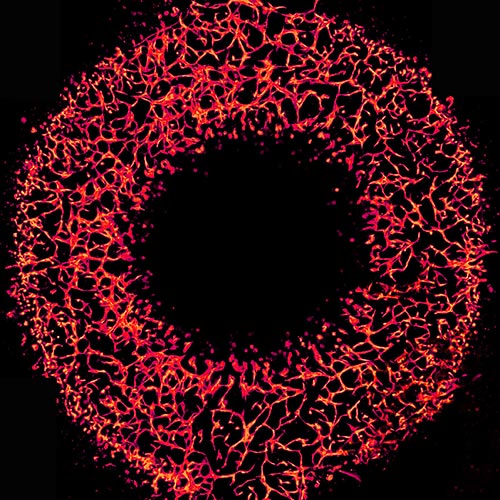 "A Microvascular Ring" - The fluorescent photograph of 3D vasculogenic network from Human Umbilical Vein Endothelial Cells inside the ring-shaped polymethylmethacrylate microfluidic chamber.
"A Microvascular Ring" - The fluorescent photograph of 3D vasculogenic network from Human Umbilical Vein Endothelial Cells inside the ring-shaped polymethylmethacrylate microfluidic chamber.
MicroTAS 2019 Conference, Basel, SWITZERLANDJoseph de Rutte,University of California, Los Angeles, USA
Show Details
Hide Details
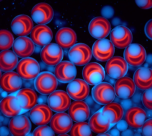 "A Cell's World": Fluorescent image of uniform droplets formed using structured microparticles. Fluorescently labeled particles are suspended in a water solution and agitated with oil and surfactant. This platform is used to encapsulate single-cells and measure their secretions.
"A Cell's World": Fluorescent image of uniform droplets formed using structured microparticles. Fluorescently labeled particles are suspended in a water solution and agitated with oil and surfactant. This platform is used to encapsulate single-cells and measure their secretions.
MicroTAS 2018 Conference, Kaohsiung, TAIWANNam-Trung Nguyen, Griffith University, Australia
Show Details
Hide Details
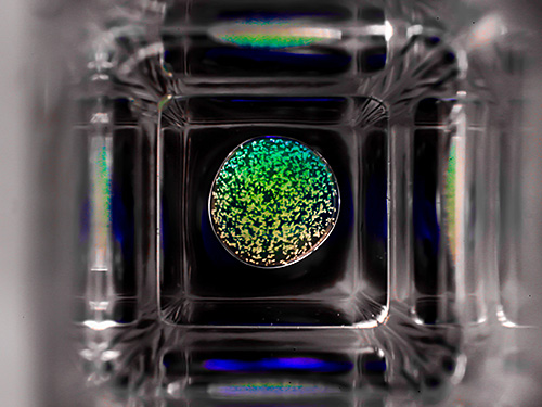
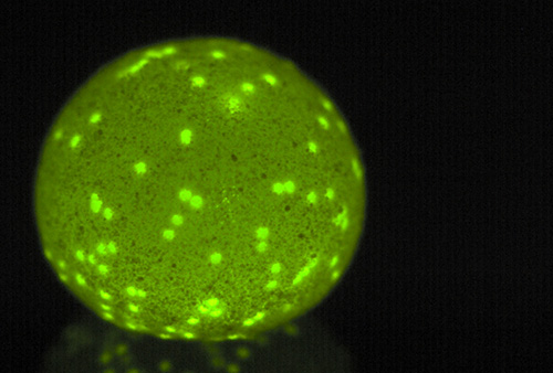 "The Green Planet". An image of a floating liquid marble, decorated with green fluorescent beads. The image was taken with a colour USB camera. The liquid marble is made of a water droplet containing green fluorescent beads and coated with Teflon powder.
"The Green Planet". An image of a floating liquid marble, decorated with green fluorescent beads. The image was taken with a colour USB camera. The liquid marble is made of a water droplet containing green fluorescent beads and coated with Teflon powder.
MicroTAS 2017 Conference, Savannah, Georgia, USAMaria Cristina Letizia, EPFL, SWITZERLAND
Show Details
Hide Details
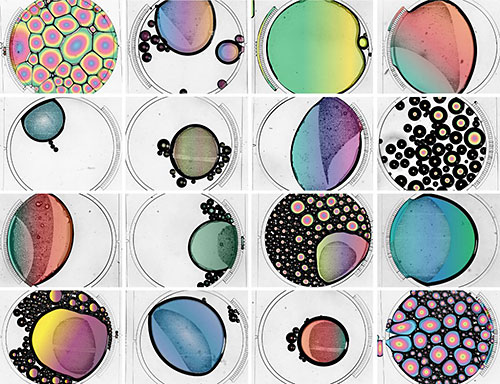 This image was obtained during the filling of microfluidic chambers with Pluronic F-127 with the purpose of functionalizing the chip.
This image was obtained during the filling of microfluidic chambers with Pluronic F-127 with the purpose of functionalizing the chip.
MicroTAS 2016 Conference, Dublin, IRELANDVaibhav Jain, Purdue University, USA
Show Details
Hide Details
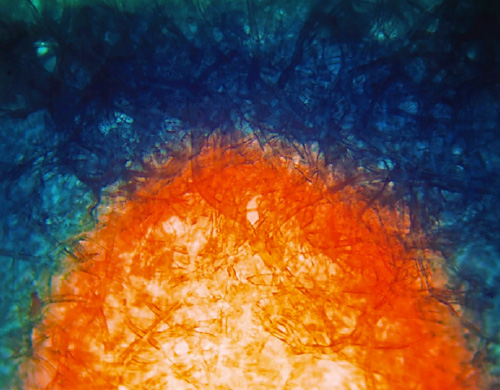 The Rising Sun by Vaibhav Jain. Dark field micrograph of sweat advancing in a paper channel dyed with Cobalt chloride dihydrate (Bluish), changing it to cobalt chloride hexahydrate (Reddish orange).
The Rising Sun by Vaibhav Jain. Dark field micrograph of sweat advancing in a paper channel dyed with Cobalt chloride dihydrate (Bluish), changing it to cobalt chloride hexahydrate (Reddish orange).
MicroTAS 2015 Conference, Gyeongju, KOREAMatteo Cornaglia, EPFL, SWITZERLAND
Show Details
Hide Details
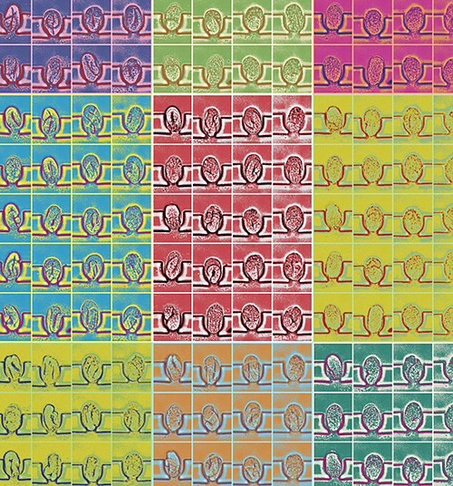 Through Warhol's Eyepiece by Matteo Cornaglia. Created by on-chip multi-dimensional imaging of C.elegans embryogenesis as observed through an Andy Warhol microscope, equipped with a 63x oil immersion objective and a pop art optical filter.
Through Warhol's Eyepiece by Matteo Cornaglia. Created by on-chip multi-dimensional imaging of C.elegans embryogenesis as observed through an Andy Warhol microscope, equipped with a 63x oil immersion objective and a pop art optical filter.
MicroTAS 2014 Conference, San Antonio, Texas, USADavid Castro, King Abdullah University of Science and Technology, SAUDI ARABIA
Show Details
Hide Details
 Al Kora, The Sphere by David Castro. Top view of a rotating ~40 uL aqueous droplet, suspended at the interface between two fluids (perfluorohexane and mineral oil) inside a square cuvette. The droplet contains an assay of functionalized latex beads, agglutinating in the presence of human C-reactive protein. Photograph by David Castro and David Conchouso.
Al Kora, The Sphere by David Castro. Top view of a rotating ~40 uL aqueous droplet, suspended at the interface between two fluids (perfluorohexane and mineral oil) inside a square cuvette. The droplet contains an assay of functionalized latex beads, agglutinating in the presence of human C-reactive protein. Photograph by David Castro and David Conchouso.
MicroTAS 2013 Conference, Freiburg, GERMANYYe Wang, Eindhoven University, THE NETHERLANDS
Show Details
Hide Details
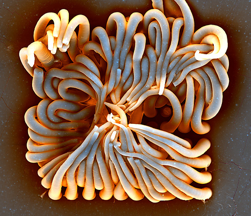 Structure and rheology of complex fluids by Ye Wang. Artificial cilia (microhairs) made with Polydimethylsiloxane and magnetic nanoparticles using a glass mold made by femtolaser modification and hydrofluoric acid etching.
Structure and rheology of complex fluids by Ye Wang. Artificial cilia (microhairs) made with Polydimethylsiloxane and magnetic nanoparticles using a glass mold made by femtolaser modification and hydrofluoric acid etching.
MicroTAS 2012 Conference, Okinawa JAPANYi Zhang, Johns Hopkins University, USA
Show Details
Hide Details
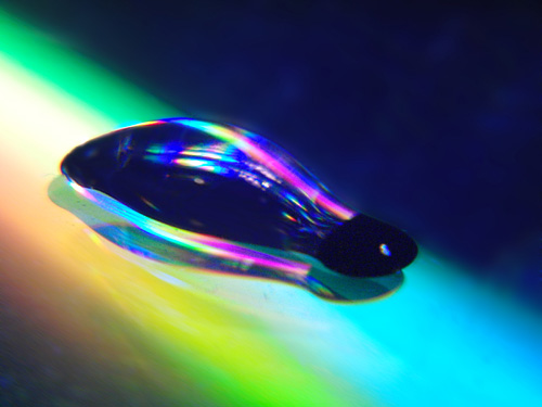 Stretching the Rainbow by Yi Zhang. A tiny water droplet held in place by surface energy traps and stretched using a magnetic field to pull on magnetic particles within it.
Stretching the Rainbow by Yi Zhang. A tiny water droplet held in place by surface energy traps and stretched using a magnetic field to pull on magnetic particles within it.
MicroTAS 2011 Conference, Seattle, Washington, USADong Jin Shin, Johns Hopkins University, USA
Show Details
Hide Details
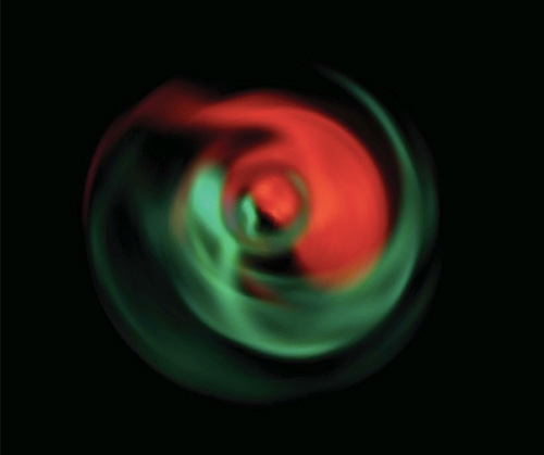 Yin and Yang in a Droplet by Dong Jin Shin. A snapshot of the mixing of two types of quantum dot solutions inside a sessile droplet driven by a microfluidic magnetic gyromixer.
Yin and Yang in a Droplet by Dong Jin Shin. A snapshot of the mixing of two types of quantum dot solutions inside a sessile droplet driven by a microfluidic magnetic gyromixer.
MicroTAS 2010 Conference, Groningen, THE NETHERLANDSNicholas Gunn, University of California, Irvine, USA
Show Details
Hide Details
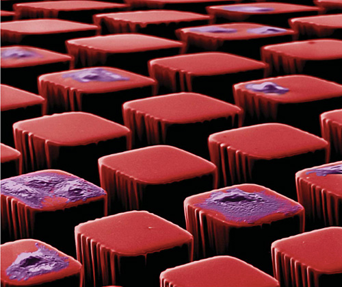 Cell Block 9 by Nicolas Gunn. A colorized SEM micrograph showing fibroblast cells cultured on microscale pedestals.
Cell Block 9 by Nicolas Gunn. A colorized SEM micrograph showing fibroblast cells cultured on microscale pedestals.
MicroTAS 2009 Conference, Jeju, KOREAJungkwun Kim, University at Buffalo, The State University of New York, USA
Show Details
Hide Details
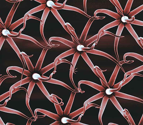 Micro world: united we stand by Jungkwun Kim and Yong Kyu Yoon. SU-8 shapes defined by multi- directional ultraviolet lithography, where glass with a pre-patterned chromium layer is used as a photomask as well as a substrate.
Micro world: united we stand by Jungkwun Kim and Yong Kyu Yoon. SU-8 shapes defined by multi- directional ultraviolet lithography, where glass with a pre-patterned chromium layer is used as a photomask as well as a substrate.
MicroTAS 2008 Conference, San Diego, California, USAYu-Wen Huang, Texas A&M University, USA
Show Details
Hide Details
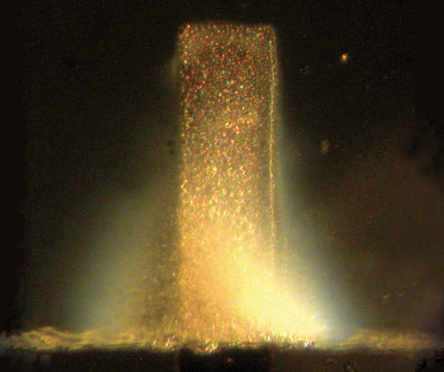 City Lights by Yu-Wen Huang. The image is a micro- graph of a solution of unlabelled double-stranded DNA, electrophoretically concentrated near a 50 mm-wide electrode.
City Lights by Yu-Wen Huang. The image is a micro- graph of a solution of unlabelled double-stranded DNA, electrophoretically concentrated near a 50 mm-wide electrode.